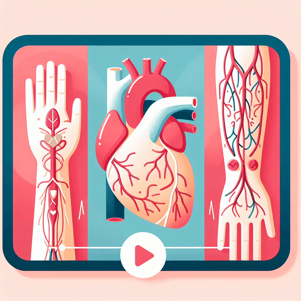
Myocardial Infarction & Peripheral Vascular Diseases
Concept-focused guide for Myocardial Infarction & Peripheral Vascular Diseases.
~6 min read
🎓 Listen to Professor Narration
Too lazy to read? Let our AI professor teach you this topic in a conversational, engaging style.
Overview
Welcome! In this session, we’ll unravel the essential concepts behind myocardial infarction (MI) and peripheral vascular diseases (PVD)—two cornerstone topics in cardiovascular nursing. You’ll learn how to interpret enzyme markers, ECG changes, and clinical findings, as well as how to prioritize nursing interventions and patient education. By the end, you’ll be ready to tackle questions about diagnosis, treatment, complications, and holistic care with confidence and clinical insight.
Concept-by-Concept Deep Dive
Cardiac Biomarkers and Diagnosis of Myocardial Infarction
Cardiac biomarkers are substances released into the blood when the heart muscle is damaged, most notably during an MI. Understanding these lab values is crucial for diagnosis.
Key Enzymes and Their Patterns
- Troponin (T and I): The gold standard for MI diagnosis. These proteins rise within hours of injury and remain elevated for days.
- Creatine Kinase-MB (CK-MB): Rises shortly after MI but returns to normal faster than troponins.
- Myoglobin: Rises quickly but is less specific to cardiac tissue.
Interpretation Steps
- Timing: Know when each marker rises and falls post-injury.
- Specificity: Troponins are most specific to cardiac damage.
- Trend Analysis: Serial measurements help confirm MI.
Common Misconceptions
- Believing all elevated enzymes mean MI—other conditions can elevate some markers.
- Overlooking timing—early or late sampling can miss the diagnostic window.
ECG Changes and Their Clinical Implications
The electrocardiogram (ECG) provides rapid, non-invasive insight into cardiac health, especially during acute events.
Key Patterns
- ST Elevation: Indicates acute injury to the myocardium, often seen in ST-Elevation Myocardial Infarction (STEMI).
- ST Depression/T-Wave Inversion: Suggests ischemia, not always full-thickness MI.
- Q Waves: Signify tissue necrosis from a completed infarct.
Clinical Steps
- Recognize the pattern (e.g., ST elevation).
- Correlate with symptoms and biomarkers for diagnosis.
- Initiate appropriate interventions rapidly.
Misconceptions
- Assuming all chest pain yields classic ECG changes—not all MI patients show textbook findings.
- Confusing ST elevation with other conditions (e.g., pericarditis).
Peripheral Vascular Disease: Signs, Assessment, and Management
PVD encompasses disorders of blood flow in arteries or veins outside the heart and brain, often leading to ischemic symptoms in the extremities.
Typical Symptoms and Findings
- Claudication: Pain in legs with activity, relieved by rest.
- Arterial Insufficiency: Cool, pale extremities, weak pulses, delayed capillary refill, ulcers on pressure points.
- Venous Insufficiency: Edema, brown discoloration, ulcers near ankles.
Physical Assessment
- Inspect for color, temperature, wounds.
- Palpate pulses; use Doppler if needed.
- Measure Ankle-Brachial Index (ABI): Ratio of ankle to brachial systolic BP.
🔒 Continue Reading with Premium
Unlock the full vlog content, professor narration, and all additional sections with a one-time premium upgrade.
One-time payment • Lifetime access • Support development
Join us to receive notifications about our new vlogs/quizzes by subscribing here!